ANATOMY
|
The outer ear consists of two primary components, the pinna and the external auditory meatus, or, the ear canal. The pinna, primarily composed of cartilage and ligaments, is the visible, flap-like structure of the hearing mechanism that is attached to the side of the head with ligaments and muscles. Unique to each individual, the pinna is flexible and covered in skin, or, epidermis and has several depressions and ridges (Szymanski). Not only this, but it also consists of some extrinsic as well as intrinsic muscles. Although of little use to humans, these muscles play a massive role in the keen sense of localization in animals (Musiek). There are eleven primary landmarks of the pinna that create the ridges and depressions making them unique to each individual. As seen on the diagram to the right, the primary ridges of the pinna include the helix, or, the outer rim of the pinna located superiorly (above) to all other landmarks. The crus of the helix runs medially, or, towards the center of the pinna and meets the concha cymba. The anti-helix, located just above the concha cymba, is the ridged landmark parallel to the helix. Made of cartilage, the tragus—located closest to the attachment point of the pinna—is the lateral opening of the ear canal. The anti-tragus is located opposite of the tragus, and hovers over the intertragal notch, which is the inferior-most (towards the bottom) depression of the pinna. The other depressions include the scaphoid fossa, which lies between the helix and anti-helix, the triangular fossa which is located laterally (towards the side) of the scaphoid fossa, and the concha cave, which is the entry to the ear canal. The lobe being the most inferior landmark of them all, is a fatty tissue covered in epidermis.
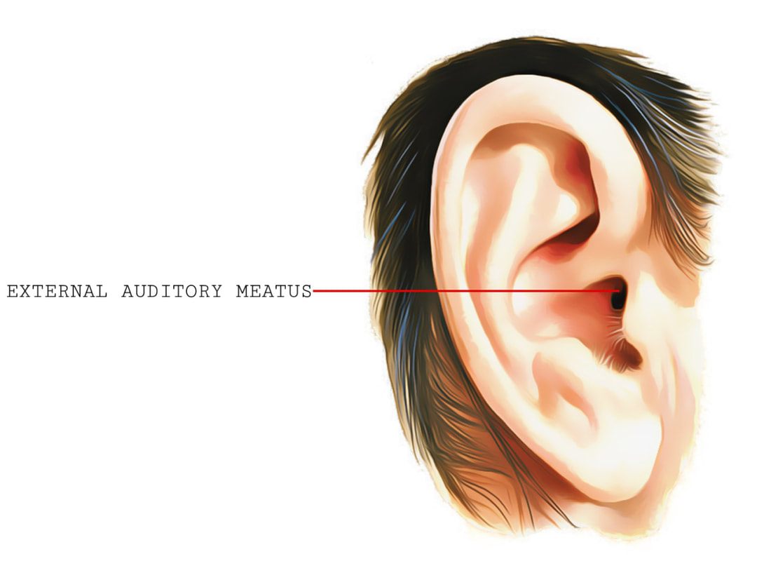
The external auditory meatus, or, the ear canal is also lined with epidermis that covers the length of the canal and continues to the outermost layer of the tympanic membrane (eardrum), which separates the middle ear from the outer ear. It consists of both bony and cartilage portions, and is shaped like a tube, with one open and one closed end. Due to the ear canal's configuration, it generates an ear canal resonance, determined by the dimensions of one's ear canal. By resonance, I mean the enhancement of an acoustic signal, which is important for an individual's natural speech perception. Without this ear canal response, patients have complained of sound perception sounding strange or "tinny," such as a ringing sound (Musiek). The cartilaginous portion of the external auditory meatus is lined with sebacious and ceruminous glands, which secrete a brown, wax-like substance called cerumen, or what we commonly refer to as ear wax. Not only does it contain these glands, but also hairs that protect the ear from foreign bodies, such as insects or dust, from entering the ear canal (How). |

|
PHYSIOLOGY

|
As we discussed above, the muscles and ligaments of the pinna are vestigial structures in humans, however, the shape of the pinna does act as a sound collection device. It absorbs and funnels sound vibrations and directs them through the external auditory meatus, and also allows us to know the source and direction of incoming sounds. As we discussed above, the external auditory meatus is responsible for reinforcing the acoustic energy that enters it and directing that energy to the middle ear, boosting the level of frequencies that are taken in from the outer ear. Due to its closed-end configuration, the sound travels to the tympanic membrane, which allows some of the acoustic energy to travel into the middle ear and reflects the rest back into the ear canal itself, creating a damping effect.
|
