ANATOMY
|
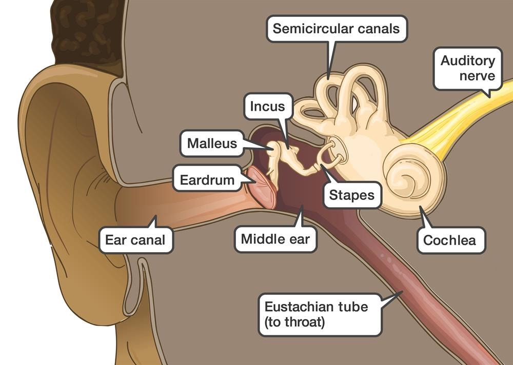
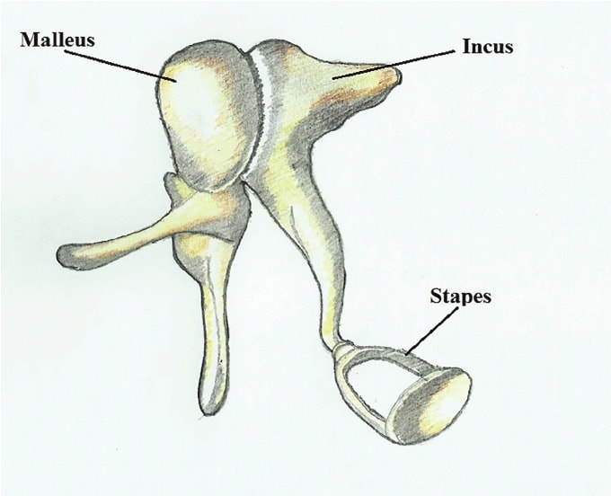
The middle ear is an air-filled, membrane lined space situated between the inner and outer ear, and it's encased within the temporal portion of the skull. The portion of the ear internal to the tympanic membrane (eardrum), the primary landmarks of the middle ear include the Eustachian tube, the ossicle bones, the middle ear muscles, and the oval and round windows. The middle ear lies within the temporal bone, and it extends from the tympanic membrane to the lateral (away from the mid-line) wall of the inner ear. The tympanic membrane is made up of thin connective tissue membrane covered by epidermis, and it separates the outer ear from the middle ear (Musiek). The Eustachian tube runs from the anterior (front-most) wall of the middle ear cavity and connects to the posterior wall of the nasopharynx, which is the upper part of the throat. It acts as a pressure equalizer, ensuring that the pressure in the middle ear is equal to that of the pressure outside of the body (Szymanski). The ossicle bones play a major role in the transmission of sound through the middle ear, and they're formed together in what is known as the ossicular chain. The three smallest bones in the human body, the malleus, incus, and stapes, form a bridge from the tympanic membrane to the inner ear. The most lateral of the three, the malleus, is a club shaped bone with three primary parts: the head, neck and processes. The malleus is about nine millimeters in length, and its head contains an articular surface where it can connect to the incus beside it. The bone's lateral and anterior processes protrude just below the head and neck and the handle is located most inferior (low) on the malleus. The neck is also the connection to the tensor tympani muscle, which aids in dampening loud noises. The articular surface on the malleus attaches to that of the incus', which also has three primary landmarks: the body, short process, and long process. The short process extends towards the back of the middle ear wall and acts as a pivot point for rotation. The long process connects the rest of the bone to the stapes, by the way of the lenticular process which is located most inferior. The stapedius muscle connects to the stapes by its head and the stapes is connected to the incus by its neck. The stapes innervates the cochlea in the inner ear by the way of the annular footplate, or, the rounded flat portion at its bottom (Musiek).
|
PHYSIOLOGY
|
The primary function of the middle ear is to transmit acoustic energy from the outer ear to the inner ear, which can then be directed to the brain. When sound first enters the external auditory meatus, or, the ear canal, it is funneled towards the tympanic membrane in the form of sound vibrations. These sound waves strike the tympanic membrane, causing it to vibrate and subsequently vibrate the ossicle bones: the malleus, the incus, and the stapes. The ossicles play a major role in the transmission of sound, as the vibration from the tympanic membrane to each of the three bones forces the medial-most (central) bone, the stapes, in a piston-like fashion in and out of the oval window. The motion of the ossicles amplifies the sound, and the vibrations are sent into the fluid-filled cochlea. An important feature of the cochlea, the round window, juts in and out along with the movement of the stapes into the oval window, acting as a pressure equalizer and allowing pressure transmitted into the cochlea to be dispersed from the opposite side of the basilar membrane, as seen in the image here. Not only this, but the round window allows for cochlear fluid to move efficiently; a traveling wave forms on the basilar membrane as a result of the rippling cochlea fluid, and the hair cells embedded in it move up and down with it (How). The hair-like projections atop the hair cells, or stereocillia, also move and bend, opening channels and allowing for chemicals to rush into the cells, creating an electrical impulse (How). This is then directed through the auditory nerve and into the brain, where it can be recognized as sound.
|

|
